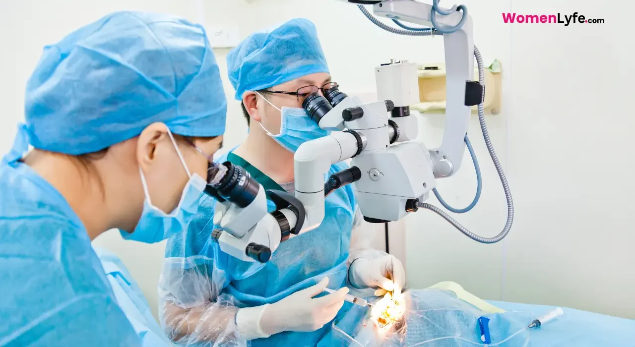
Cataract surgery is one of the most popular and commonly performed procedures in the world. The vast majority of patients have excellent outcomes with few complications.
Here are the numbers:
- By age 80, over half of all Americans have cataracts.
- Close to 4 million cataract surgeries are performed in the U.S. every year.
- Over 90% of patients have 20/20 vision with glasses after surgery, although those with other eye conditions may not do as well, including those with glaucoma, a progressive disease typically associated with elevated pressure within the eye; diabetic retinopathy, which ultimately can cause leakage in the retinal tissues; and macular degeneration, a disease that is typically related to age.
- The rate of post-surgery infection from endophthalmitis is less than 0.1%.
As ophthalmologists who have performed thousands of these procedures, we know that many patients have misconceptions about both cataracts and the surgery. For example, some think a cataract is a growth on the eye’s surface.
We like to compare a cataract with the frosted glass of a bathroom window, where light can be transmitted but details cannot. Or when turbulence from a storm causes normally clear water in the ocean to become murky. In much the same way, the eye’s once transparent lens becomes cloudy.After surgery, there’s no bending, inversions, lifting or straining, high-impact activities or eye makeup for one to two weeks or until the doctor says it’s OK.
About the Cataract surgery
Cataract surgery removes the clouded lens of the eye and replaces it with a new, clear lens to restore your vision. Most patients report the procedure is painless.
It’s typically an elective surgery that is performed on an outpatient basis. The patient is often awake, under local anesthesia, with sedation similar to that used for dental procedures. We like to say patients receive the equivalent of three margaritas in their IV.
Numbing drops are then applied to the eye’s surface, along with an anesthetic inside the eye. Patients with claustrophobia, or movement disorders such as Parkinson’s disease, may not be suitable candidates for awake surgeries and require general anesthesia.
Before surgery, patients receive dilating drops to make the pupil as large as possible. The surgeon makes a tiny incision, usually with a small pointed scalpel, between the clear and white part of the eye to gain access to the lens capsule, a thin membrane similar in thickness to a plastic produce bag at the grocery store.
This capsule is suspended by small fibers called zonules, which are arranged like the springs that suspend a trampoline from a frame. The surgeon then creates a small opening in the capsule, called a capsulotomy, to gain access to the cataract. The cataract is then broken into smaller parts so they are removable through the small incision.
This is similar to a tiny jackhammer, breaking the large lens into smaller pieces for removal. That sounds scary, but it’s painless. Ultrasound emulsifies the lens and vacuum power then aspirates it from the eye.
Laser-assisted cataract surgery has been found to have similar outcomes to traditional cataract surgery.
Complications are rare
Serious complications, such as postoperative infection, bleeding in the eye or a postoperative retinal detachment are rare; they occur in approximately 1 in 1,000 cases. But even in many of these situations, appropriate management can salvage useful vision.
Capsular complications deserve additional discussion. According to some studies, they occur in up to 2% of cases. If a hole or tear of the posterior capsule is encountered during cataract surgery, the clear gel in the vitreous – the back chamber of the eye – may be displaced into the front chamber of the eye.
If that happens, the gel must be removed at the time of the cataract surgery. This will reduce the likelihood of additional postoperative complications, but those who have the procedure, known as a vitrectomy, have an increased risk for additional complications, including postoperative infections and postoperative swelling.
Also read
After Cataract surgery
Patients usually go home right after the procedure. Most surgery centers require that the patient have someone drive them home, more for the anesthesia rather than the surgery. Patients begin applying postoperative drops that same day and must wear an eye shield at bedtime for a few weeks after surgery.
Patients should keep the eye clean and avoid exposure to dust, debris and water. They should try not to bend over and should avoid heavy lifting or straining in the first week or so after surgery. Lifting or straining can cause a surge of blood pressure to the face and eye. Known as a choroidal hemorrhage, it can lead to bleeding into the wall of the eye and be devastating to vision.
Things that cause only moderate increases in heart rate such as walking are OK. Routine postoperative examinations are usually completed the day after surgery, about a week after surgery and about a month after surgery.Light and UV exposure, coupled with time, causes the lens of the eye to become increasingly cloudy.
A choice of lens: Intraocular Lens
The plastic lens used to replace the cataract, or intraocular lens, requires careful sizing for optimal results and a nuanced discussion between patient and surgeon.
Early intraocular lens technologies were monofocal, and most patients with these lenses chose distance correction and used reading glasses for near tasks. This is still the preferred approach for approximately 90% of patients having cataract surgery today.
Recent advances have led to intraocular lenses that offer multifocality – the opportunity to have near as well as distance vision, without glasses. Some multifocal lenses are even in the trifocal category, which includes distance, near, and intermediate vision, the latter of which in recent years has become very important for computer and phone use.
Most patients with these advanced technology multifocal lenses are happy with them. However, a small percentage of patients with multifocal lenses can be so bothered by visual disturbances – notably night glare and halos around light sources in the dark – that they request removal of the multifocal lens to exchange it for a standard intraocular lens. These exchanges are a reasonable option for such situations and offer relief for most affected patients.
Determining who’s an ideal candidate for a multifocal intraocular lens is an area of active research. Most clinicians would recommend against such a lens for a patient with a detail-oriented personality. Such patients tend to fixate on the shortcomings of these lenses despite their potential advantages.
As with many technologies, current generation advanced technology intraocular lenses are much better than their predecessors. Future offerings are likely to offer improved vision and fewer side effects than those available today.
But these newer lenses are often not reimbursed by insurance companies and often entail substantial out-of-pocket costs for patients.
Deciding on what type of lens is best for you can be complicated. Fortunately, except in unusual circumstances, such as when a cataract develops after trauma to the eye, there is seldom a hurry for adult cataract surgery.
Allan Steigleman is an Associate Professor of Ophthalmology at the University of Florida. Elizabeth M. Hofmeister is an Associate Professor of Surgery at the Uniformed Services University of the Health Sciences. This article is republished from The Conversation under a Creative Commons license. Read the original article.


